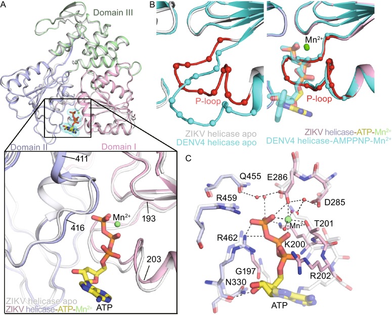
Figure 2.
Structure of the ZIKV helicase in complex with ATP/Mn 2+. (A) Overall fold of ZIKV helicases, with cartoon representation of apo form (white) overlaid to the complex (the three domains are colored respectively). The ATP is drawn as sticks and mesh; Mn2+ as green sphere. A detailed comparison for the ATP binding sites of the two structures is depicted in the zoomed view below. (B) A close-up view of the NTPase active site. P-loops are represented by superimposition of the structures of ZIKV (white, with the P-loop highlighted in red) and DENV4 (cyan) apo-helicases in the left panel and their complexes in the right panel. The DENV4 helicase complex was bound to AMPPNP and Mn2+ (PDB code 2JLR). (C) Interactions at NTPase active site by superposition of the ZIKV helicase complexed with ATP and Mn2+ (solid) with its apo enzyme (semitransparent, PDB code 5JMT)

