
Figure 2.
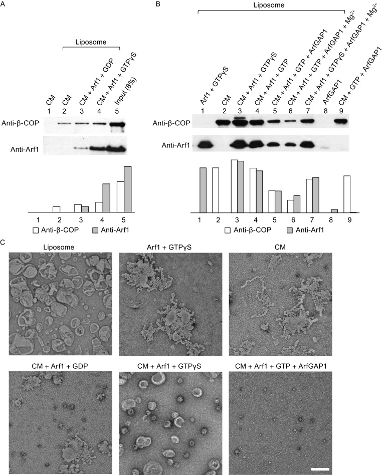
Membrane binding and vesiculation assays. (A and B) Membrane binding co-sedimentation assays (See Materials and methods). The amount of co-sedimented coatomer (β-COP) and ARF1 is also quantified in lower panels. (C) Negative-stain electron microscopy of liposomes incubated with coatomer and other factors. Scale bar, 200 nm. CM, human coatomer; ARF1, N-myristoylated human ARF1; ARFGAP1, rat ARFGAP1