Figure 4.

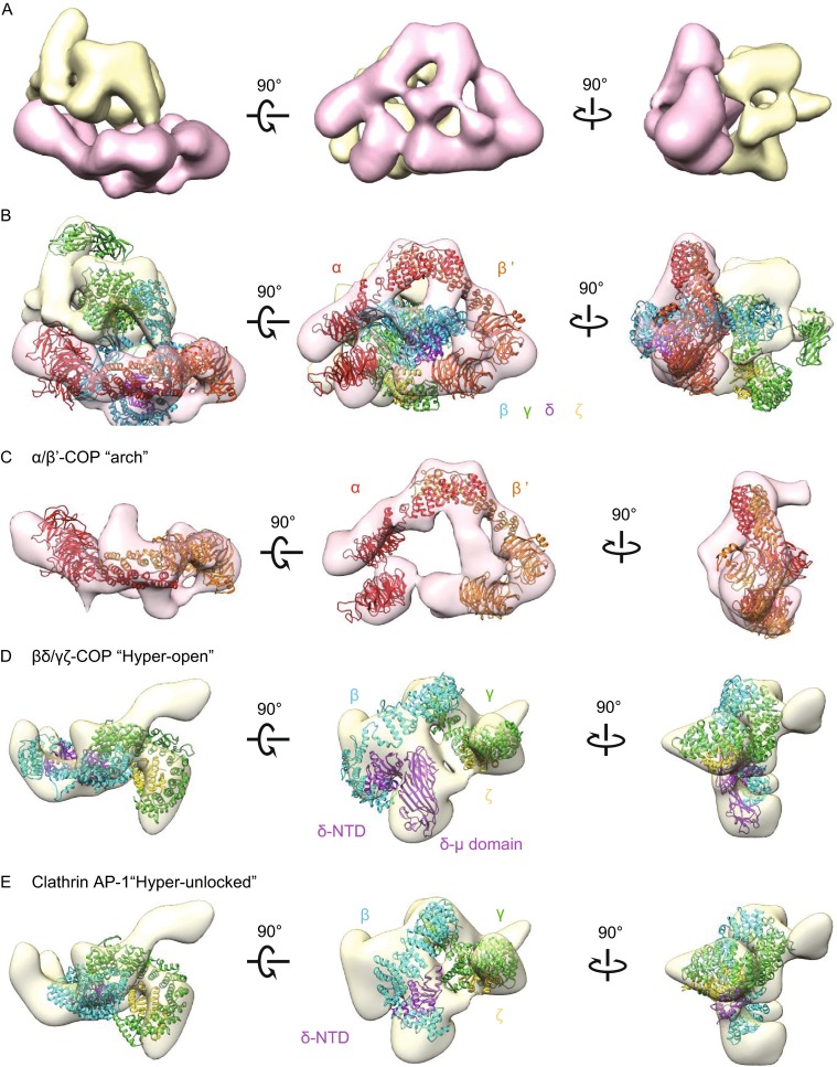
3D reconstruction of the recombinant human coatomer in its soluble form. (A) Three views of surface-rendered density map of coatomer obtained by single-particle reconstruction. The map is segmented using UCSF Chimera, grouped, and colored with pink for the arch-shape portion and yellow for the more globular portion. (B) Fitting the membrane-bound model of coatomer (PDB ID: 5A1U) into the EM map. (C) Fitting the B-subcomplex αβ′-COP heterodimer into the “focus” refined arch-shape density map. (D) Fitting the F-subcomplex βδ/γζ-COP into the “focus” refined globular density map. (E) Fitting the homologue model of F-subcomplex in “hyper-unlocked” form into the “focus” refined globular density map. α-, β′-COP are colored in red and orange. β-, δ-, γ-, and ζ-COP and their homologues are colored in cyan, purple, green, and yellow respectively
