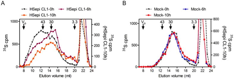
Figure 3. Pulse-chase labeling of HS in HEK293 cells.
Gel chromatography on Superose-6 of HS samples isolated from Hsepi Clone 1 (A) and Mock cells (B) after metabolic 35S pulse-labeling for 30 min followed by chase-incubation for the indicated periods of time. Elution positions of polysaccharide standards (43, 30 and 3.3 kDa, respectively), are indicated. The peaks eluted after 20 ml are degradation products of chondroitin sulfates.

