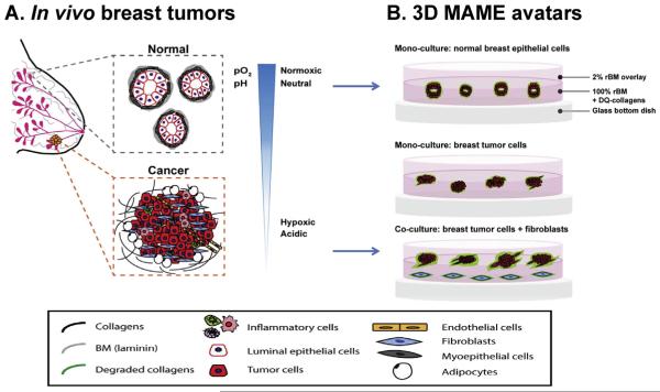
Fig. 2.
Schematic illustrations of normal versus cancerous human breast cells within the in vivo breast microenvironment (A) and as modeled in our 3D MAME avatars (B). In vivo, normal human breast ducts, comprised of myoepithelial cells and luminal epithelial cells (i.e., epithelial cells lining the lumen) (A, gray dashed box), grow in a controlled normoxic and neutral pH microenvironment. In contrast, breast cancer cells interact with adjacent and infiltrating stromal cells (e.g., fibroblasts, inflammatory cells, endothelial cells, and adipocytes) (A, orange dashed box) and grow irregularly in a hypoxic and acidic tumor microenvironment. Our 3D MAME avatars are designed to mimic in vivo normal and cancerous breast tissues (B). Normal epithelial cells grown in 3D MAME cultures form acinar structures with lumens resembling normal breast tissue (B, top image) and exhibit a basal level of DQ-collagen IV degradation (green). In comparison, breast ductal carcinoma in situ cells grown in 3D MAME cultures form larger, irregular and dysplastic structures (B, middle image). Co-cultures of breast carcinoma cells and fibroblasts result in an invasive morphology and increased degradation of DQ-collagen IV (B, bottom image). A was modified from Nelson and Bissell, 2005. Seminars in Cancer Biology [80].

