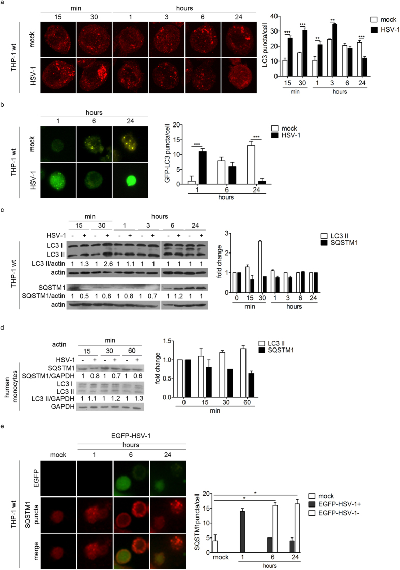
Figure 1. HSV-1 transiently induces autophagy during the early phases of the infection.
(a) Representative confocal images of THP-1 cells mock-infected or infected with HSV-1, collected at different times and stained for endogenous LC3. Quantification of LC3 puncta was performed using Imaris software; 50 cells were considered for each sample. (b) Representative images obtained using an epifluorescence microscope of THP-1 cells transiently transfected with GFP-LC3 plasmid for 6 h, then mock-infected or infected with HSV-1 and fixed at different times. GFP-LC3 puncta were quantified by counting them and 50 cells were considered for each sample. (c) Immunoblotting of LC3 and SQSTM1 in THP-1 cells infected or mock-infected at the indicated times. The densitometric analysis of the LC3II/actin and SQSTM1/actin ratios is reported. (d) Immunoblotting of LC3 and SQSTM1 in human monocytes infected or mock-infected at the indicated times. The results shown are representative of three independent donors. (e) Immunofluorescence images of THP-1 cells infected with EGFP-HSV-1 and stained to identify SQSTM1. Quantification of SQSTM1 puncta in cells expressing EGFP (EGFP-HSV-1 +) or not (EGFP-HSV-1 −). *p < 0.05; **p < 0.01; ***p < 0.001.

