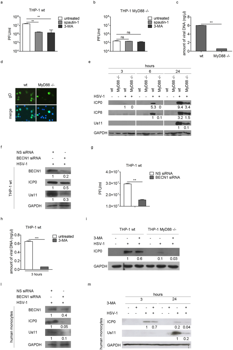
Figure 4. Induction of autophagy mediated by HSV-1 has a proviral role.
Viral production was quantified in wt (**p < 0.01) (a) and MyD88−/− THP-1 cells (b) pre-treated with spautin-1 or 3-MA and infected with HSV-1 in the presence of the inhibitors for 24 h. (c) Amount of viral DNA in wt and MyD88−/− THP-1 cells was determined by real time PCR (**p < 0.01). (d) Immunofluorescence analysis showing gD-positive wt and MyD88−/− THP-1 cells 24 h pi. Hoechst 33342 was used to stain the nuclei. (e) Immunoblot analysis of ICP0, ICP8 and Us11 in wt and MyD88−/− THP-1 cells infected with HSV-1. GAPDH was used as a loading control. (f) THP-1 cells were nucleofected with Beclin 1 (BECN1) siRNA for 24 h and then infected with HSV-1. ICP0 and Us11 expression and viral production (g) were quantified by immunoblot analysis and plaque assay, respectively. The amount of viral DNA (h) was analyzed in THP-1 cells pre-treated with 3-MA and infected with HSV-1 in the presence of the inhibitor for 3 h (***p < 0.001). The same experiment was carried out in wt THP-1 and MyD88−/− cells and ICP0 expression was evaluated by immunoblot (i). ICP0 and Us11 expression in human monocytes nucleofected with BECN1 siRNA for 6 h and then infected with HSV-1 for additional 24 h (l), or pre-treated with 3-MA and infected with HSV-1 in the presence of the inhibitor for 3 h and 24 h (m). The results shown are representative of three independent donors.

