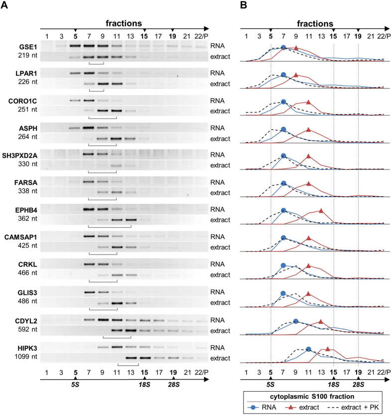
Figure 1. Evidence for distinct circRNA-protein complexes in mammalian cells.
(A) Sedimentation profiles of circRNPs/circRNAs. Cytoplasmic S100 extract and corresponding free RNA from HeLa cells were fractionated by glycerol gradient centrifugation (#1–22 from top to bottom; the last fraction containing the resuspended pellet), followed by RT-PCR analysis of 12 abundant circRNAs across the gradient (ordered from top to bottom according to their sizes; given in nucleotides). The positions of ribosomal RNA size markers are indicated (5S, 18S, and 28S), as well as the shift of the circRNA vs. circRNP peak fractions (brackets). (B) Quantitation of circRNA distribution across gradient, comparing free RNA prepared from extract (RNA, in blue), cytoplasmic S100 fraction (extract, in red), and proteinase K-treated extract (extract + PK, in dashed lines). Data from panel A (RNA/extract) and from Supplementary Fig. S1A (extract + PK) were used for quantitation.

