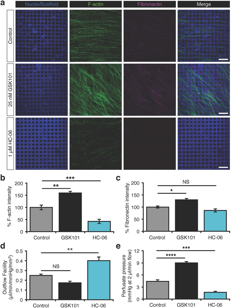
Figure 5. TRPV4 regulates the outflow facility in 3D pTM nanoscaffold models of conventional outflow together with remodeling of the cytoskeleton and extracellular matrix.
pTM-seeded SU-8 scaffolds. (a) Representative IHC images of F-actin and fibronectin following 24 hrs of perfusion. Scale bars = 30 μm. (b) Quantification of F-actin signals from (a). (c) Quantification of fibronectin signals from (a). (d) HC-06 (1 μM) increases the outflow the facility in 3D pTM cultures, whereas GSK101 (25 nM) non-significantly attenuates it (N = 5 experiments from 3 donors). (e) Relative to the vehicle control, HC-06 lowers perfusate pressure, whereas GSK101 has the opposite effect (N = 5 experiments from 3 donors).

