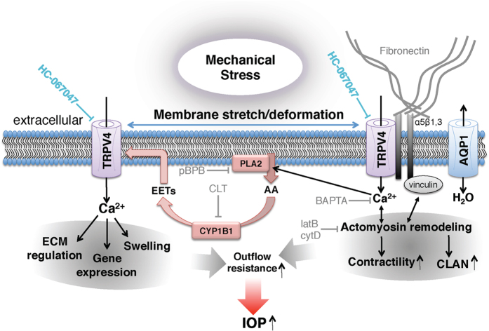
Figure 7. Model of trabecular meshwork signaling in response to mechanical stress.
Mechanical stress (e.g., pressure, swelling, and tissue distension) stretches the plasma membrane and activates TRPV4 and a Ca2+- and stretch-sensitive phospholipase A2 (PLA2). The product, arachidonic acid (AA), is a substrate for cytochrome P450, which drives synthesis of eicosanoid metabolites (EETs), the final activators of TRPV4. Stretch might activate PLA2 simultaneously with TRPV4; alternatively, stretch-induced TRPV4 activation could stimulate Ca2+-dependent PLA2s which amplify the initial TRPV4 signal (horizontal blue arrows). TRPV4 activation may be additionally augmented by TRPV1 channels and/or cell swelling, mediated through aquaporin 1 (AQP1) channels67,68,69. Stretch regulates the outflow resistance through Ca2+ -dependent actin remodeling, focal adhesion stabilization, actomyosin contractility, fibronectin production and TM gene expression. Acting in concert, these signaling components account for many known aspects of the TM response to mechanical and inflammatory challenges, including the response to the response to chronic IOP elevations.

