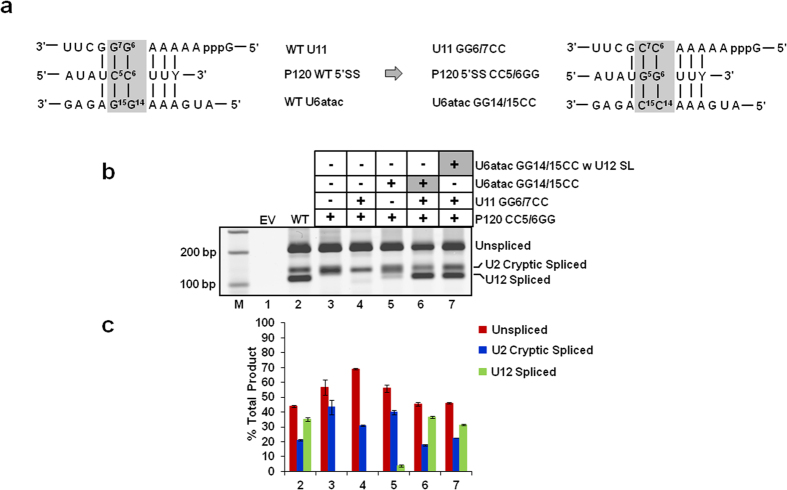
Figure 3. Effect of swapping the distal 3′ SL in U6atac snRNA with the U12 SLIII on in vivo splicing.
(a) Diagram of the 5′ splice site (SS) in vivo genetic suppression assay. The base pairing interactions between the WT (wild type) U11 snRNA and WT P120 pre-mRNA 5′ SS, and between the WT U6atac snRNA and pre-mRNA 5′SS are shown. The mutations introduced in U11 snRNA, U6atac snRNA and the P120 pre-mRNA’s 5′SS are also indicated. The boxed nucleotides (shown on the left) were mutated to their complementary nucleotides as shown in the box on the right. Both the U11 GG6/7CC and U6atac GG14/15CC mutations are required to fully suppress the effect of the 5′SS CC5/6GG mutation. (b) Splicing phenotypes of the P120 WT and P120 CC5/6GG mutant co-expressed with the indicated U11 and U6atac snRNA mutants. Total RNA was extracted from CHO cells transfected with the indicated constructs and the in vivo splicing pattern was analyzed by RT-PCR. Lane M: 100 bp ladder. U6atac GG14/15CC w U12 SL denotes the U6atac GG14/15CC snRNA construct containing the SLIII of U12 snRNA in place of the U6atac distal 3′ SL. The positions of bands corresponding to unspliced RNA, RNA spliced at the normal U12-dependent splice sites (U12 spliced) and RNA spliced at the cryptic U2-dependent splice sites (U2 cryptic) are indicated. The cryptic spliced product is a result of activation of U2-dependent cryptic splice sites in the U12-dependent intron of the P120 minigene. (c) Quantitative analysis of spliced/unspliced bands. Numbers (x-axis) correspond to the respective lanes of the gel shown in (b). Error bars represent ± SE of three experiments.

