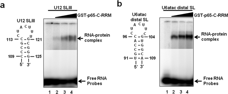
Figure 6. The C-terminal RRM of p65 binds the distal 3′ SL of U6atac snRNA.
(a) EMSA of U12 SLIII with GST-p65-C-RRM. The sequence of the WT U12 SLIII RNA oligonucleotide is shown. 32P-labeled oligonucleotide was incubated with increasing concentrations of GST-p65-C-RRM (0, 20, 40, 60 nmoles). RNA–protein complexes were separated on a 6% native polyacrylamide gel. (b) EMSA of the U6atac distal 3′ SL with GST-p65-C-RRM. The sequence of the WT U6atac distal 3′ SL RNA oligonucleotide is shown. 32P-labeled oligonucleotide was incubated with increasing concentrations of GST-p65-C-RRM (0, 20, 40, 60 nmoles). RNA–protein complexes were separated on a native 6% polyacrylamide gel. Arrows on the right denotes the position of the RNA-protein complex band and unbound RNA. The upper and lower parts of the gel represent different exposures.

