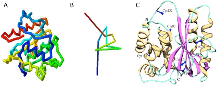
Figure 2. UCH-L1 knotted backbone.
(A) Schematic representation of the peptide backbone structure of UCH-L1. (B) A simplified schematic of UCH-L1 backbone knot. Schematics taken from [16]: Day, I.N. and Thompson, R.J. (2010) UCHL1 (PGP 9.5): neuronal biomarker and ubiquitin system protein. Prog. Neurobiol. 90, 327–362, with permission. (C) Crystal structure of UCH-L1 secondary structure highlighting the two ‘lobes’ of α-helices surrounding the β-strands in the hydrophobic core. The location of the six cysteine residues are in blue. The location of Cys90 in the catalytic triad and Cys152 in the short loop covering the active site can be observed. Schematic from [36]: Koharudin, L.M., Liu, H., Di Maio, R., Kodali, R.B., Graham, S.H. and Gronenborn, A.M. (2010) Cyclopentenone prostaglandin-induced unfolding and aggregation of the Parkinson disease-associated UCH-L1. Proc. Natl. Acad. Sci. U.S.A. 107, 6835–6840, with permission.

