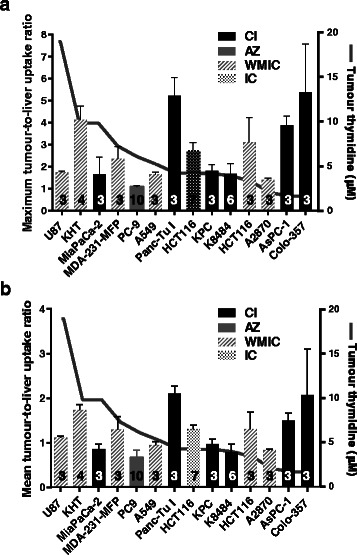
Fig. 6.

Tumour thymidine concentrations were not correlated with [18F]FLT uptake. Tumour-to-liver uptake (TTL) ratios ± SD are shown as a bar chart superimposed on a plot of the respective tumour thymidine concentrations. Left axes show uptake ratios; right axes show thymidine concentrations. a TTLmax. b TTLmean. Numbers of animals are indicated on the columns. Column fill indicates the centre supplying the tumour samples. See Additional file 1: Figure S7 and ESM tables (a), (b) and (c) for the plotted values and the median, maximum, minimum, first and third quartiles
