
Fig. 1.
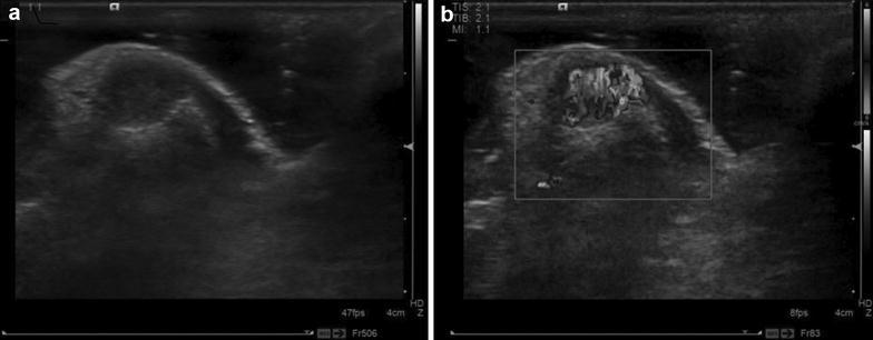
Ultrasound images of female patient (27 years old). a Two-dimensional ultrasound showed hypoechoic partial ulnar side in right thumb, with clear border and regular shape, the rear phalanx pressed to be curved; b CDFI showed rich blood flow in tumor tissues