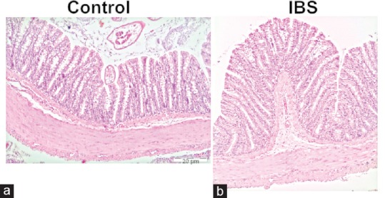
Figure 3.

Photomicrographs of hematoxylin and eosin staining within the control and IBS tissues is depicted (×10). There was no difference with regard to morphology between control (a) and IBS (b) tissues in hematoxylin and eosin staining. IBS: Irritable bowel syndrome
