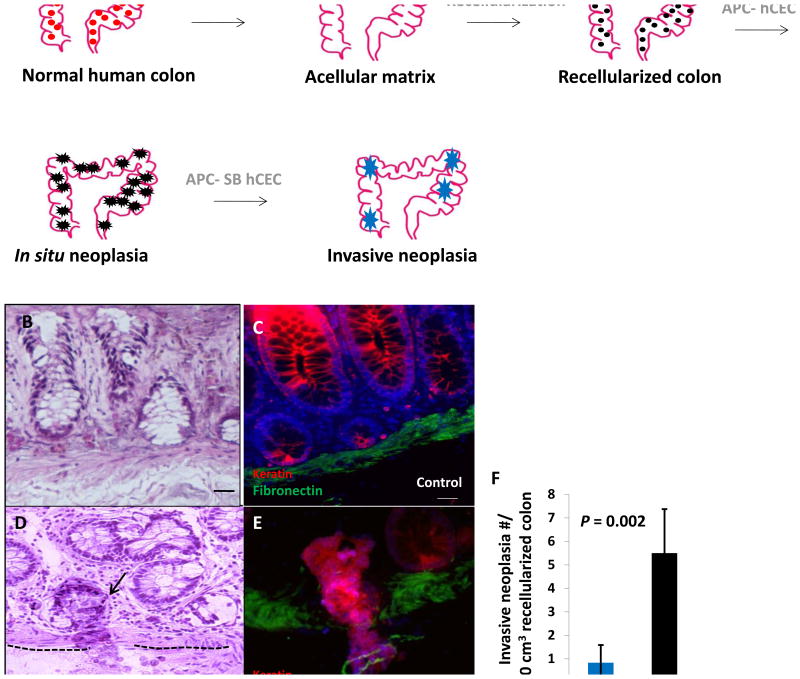
Figure 3. Invasive adenomas induced by SB mutagenesis. (A).
A schematic showing the different steps used in the creation of invasive adenomas by SB. (B,D) H+E stained images or (C,E) immunostained images of cytokeratin and fibronectin in CRC models recellularized with wild type hCECs mutagenized SB (upper panels) or APC shRNA-expressing hCECs mutagenized with SB (lower panels). Blue nuclei, DAPI; Scale bars, 50μ. (F) Quantification of invasive neoplasia in the two CRC models. All of the recellularized tissues had been cultured for 7 weeks when they were terminated for quantification of tumorigenesis. These data show that SB-mutagenesis promotes CRC progression in APC shRNA-expressing hCECs. (n=6 of independent matrix; difference analysis by 2-sided student t test). Error bars indicate S.E.M. (G-I) Dual immunostained images of cytokeratin and fibronectin showing progression from in situ neoplasia to submucosal invasion in SB-mutagenized hCECs. Blue nuclei, DAPI; Scale bars, 50μ.

