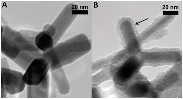
Figure 1.
Transmission electron micrograph of uncoated ZnO (A) and silica-coated ZnO (B) NPs. (Note: arrow points to the thin silica coating of approximately 5 nm in B). In both cases, the ZnO NPs have a rod-like shape with an aspect ratio of 2:1 to 8:1 (Sotiriou et al., 2014).

