
Fig. 1.
Silk string loops were applied at the aorta distally to the left renal artery and proximally to the aortic bifurcation (white arrow). The left common femoral artery was cannulated with a polyethylene-10 tube (black arrow).
Official websites use .gov
A
.gov website belongs to an official
government organization in the United States.
Secure .gov websites use HTTPS
A lock (
) or https:// means you've safely
connected to the .gov website. Share sensitive
information only on official, secure websites.
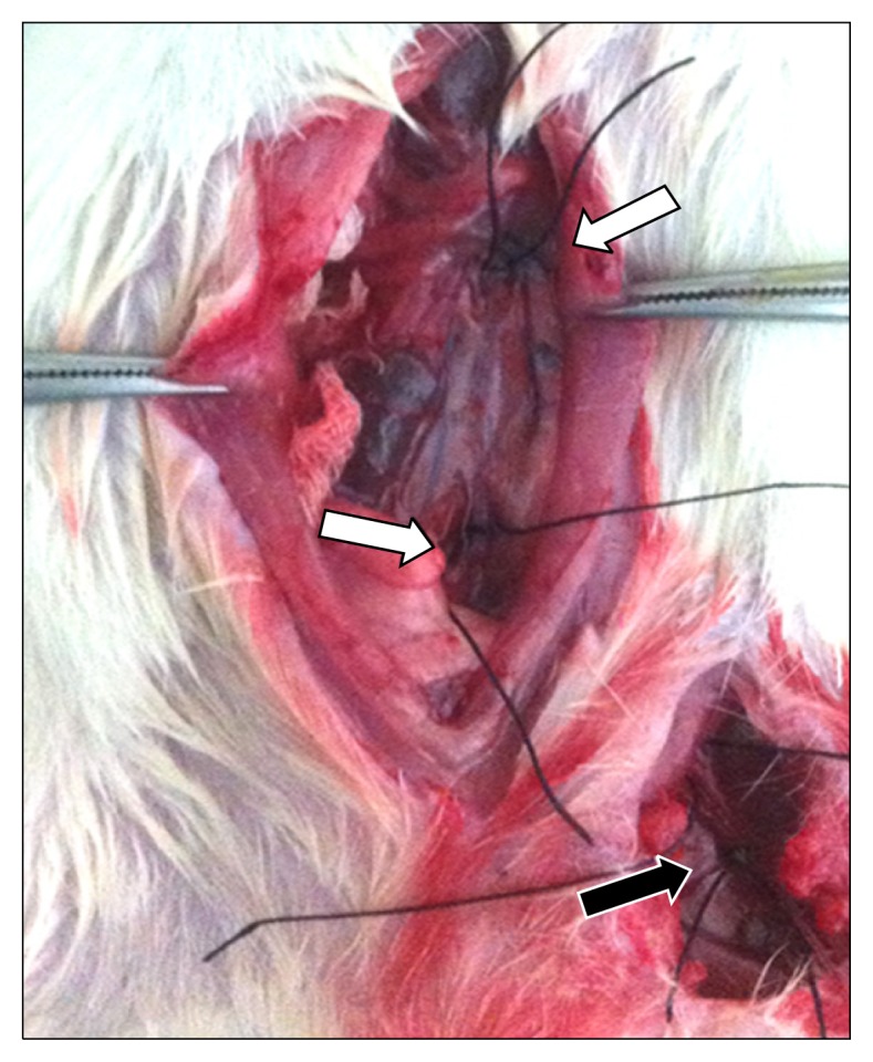
Silk string loops were applied at the aorta distally to the left renal artery and proximally to the aortic bifurcation (white arrow). The left common femoral artery was cannulated with a polyethylene-10 tube (black arrow).