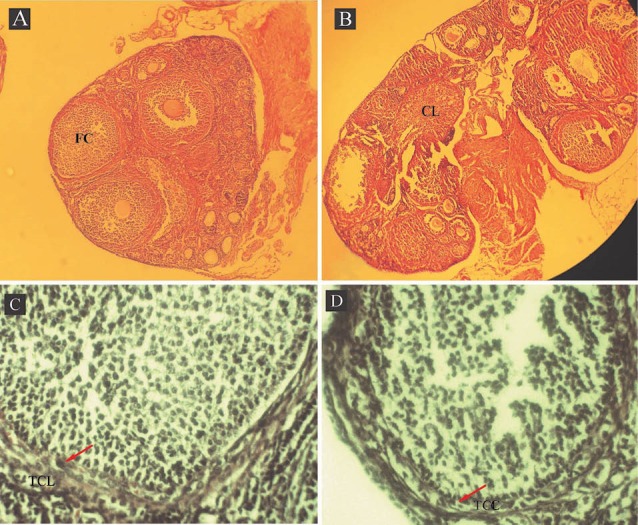
Fig. 1 .

Photomicrographs of ovaries from DHEA-treated mouse with a follicular cyst (FC) (A) and control with a corpus luteum (CL) (B). Control and DHEA-treated ovaries showed follicles at different stages of development. Representative examples of the follicular wall of a cyst (C) and a tertiary follicle (D). The arrow points to the theca cell layer (TCL). (A & B) Magnification: 1000× and (C & D) 4000×.
