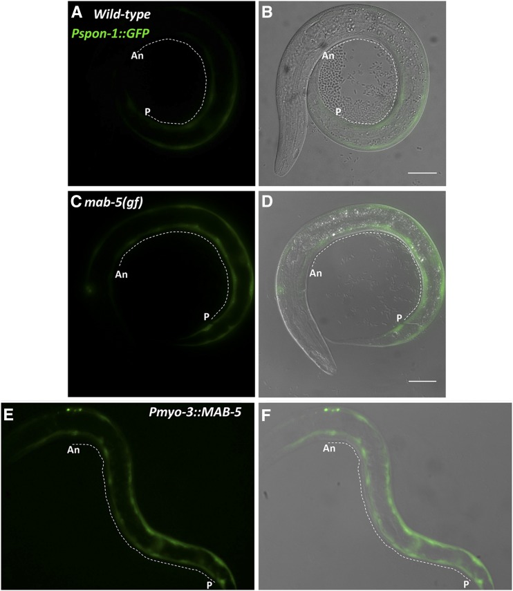
Figure 7.
Pspon-1::gfp expression in mab-5 gain of function. Fluorescent micrographs of Pspon-1::gfp-expressing L1 larvae 4–4.5 hr posthatching. Fluorescent Pspon-1::gfp (A, C, and E) and merged DIC (B, D, and F) micrographs. Dashed lines indicate regions of BWM used in line scans for the analysis in Figure 6 (An, anterior near the posterior pharyngeal bulb; P, posterior near the anus) (see Materials and Methods). Bar, 10 μm.

