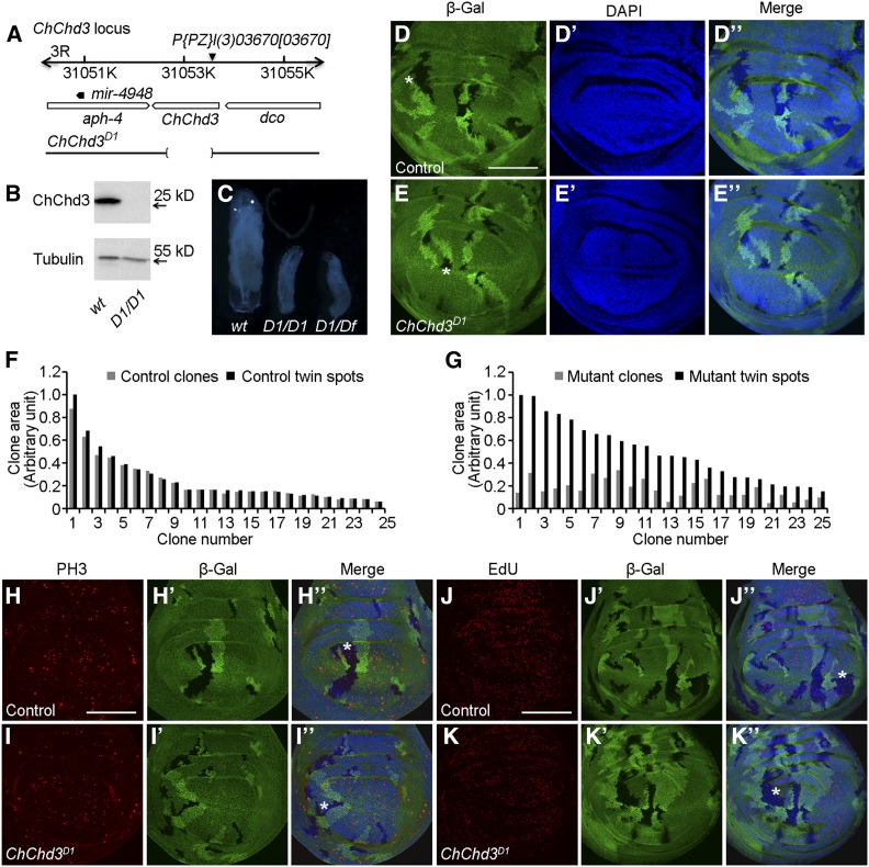
Figure 2.
Loss of function of ChChd3 results in growth defects. (A) Schematic representation of the ChChd3 locus. The deletion in ChChd3D1 mutant allele is indicated by the bracketed area. (B) Western blot on second instar larval extracts from wild-type and ChChd3D1 mutant animals. Lysates were probed with anti-ChChd3 and anti-tubulin. (C) Wild-type control and mutant larvae homozygotic for ChChd3D1 or transheterozygous for ChChd3D1 and a deficiency after 5 days of growth. D1 denotes the ChChd3D1 mutant allele. Df denotes the Df(3R)BSC749 deficiency line removing the ChChd3 locus. (D–D′′) Wing imaginal disc with control mitotic clones (lack of β-Gal, black area) and their corresponding twin spots (two copies of β-Gal, brighter area) of similar size. (E–E′′) Wing imaginal disc with ChChd3D1 homozygous mutant clones (lack of β-Gal) that are smaller than their corresponding twin spots (two copies of β-Gal). (F) Measurements of clone area for 25 pairs of control clones and their sister twin spots. (G) Measurements of clone area for 25 pairs of ChChd3D1 homozygous mutant clones and their sister twin spots. (H–I′′) Wing imaginal discs containing control (H–H′′) or ChChd3D1 homozygous mutant (I-I′′) clones stained with anti-β-Gal and anti-PH3 antibodies. ChChd3 mutant clone had reduced number of cells positive for PH3 staining. (J–K′′) Wing imaginal discs containing control (J–J′′) or ChChd3D1 homozygous mutant (K–K′′) clones stained with anti-β-Gal and labeled with EdU. ChChd3 mutant clone had reduced number of cells positive for EdU labeling. Asterisks indicate the clone area. Bars, 100 μm.

