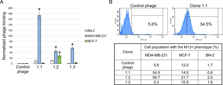
Fig 1. Relative binding of the selected bacteriophage clones to cells.
Displayed peptides 1.1 –YTYDPWLIFPAN, 1.2 –FIPFDPMSMRWE and 1.3 –SLPVYAPALTSR. (A) MDA-MB-231, MCF-7 and BN-2 cells were incubated with selected phage clones and control bacteriophage. After incubation and a series of washings, the titer of bacteriophages bound to cells was determined. Cells of untransformed human breast BN-2 primary culture were used as a negative control. The value for the binding of the control bacteriophage was used as a normalization value. Data are presented as mean ± SD. Data were statistically analyzed using one-way ANOVA with post hoc Fisher test. A p value < 0.05 was considered to be statistically significant. (B) Flow cytometry of MDA-MB-231, MCF-7 and BN-2 cells incubated with clone 1.1 and control bacteriophage. Flow cytometry was performed using mouse anti-М13 and Alexa Fluor 488 donkey anti-mouse IgG (H+L) antibodies.

