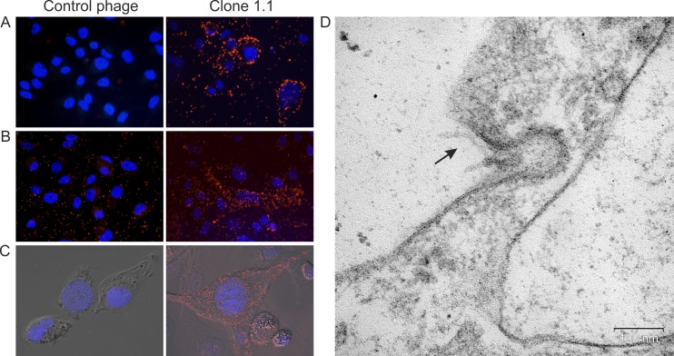
Fig 3. Displayed peptide YTYDPWLIFPAN provided penetration of phage particles into MDA-MB-231 cells.
(А) Fluorescence microscopy of MDA-MB-231 cells incubated with phage clone 1.1 and control bacteriophage at 4°C. (B) Fluorescence microscopy and (C) confocal fluorescence microscopy of MDA-MB-231 cells incubated with phage clone 1.1 and control bacteriophage at 37°C and further washes from bacteriophages bound to the surface. Fluorescence microscopy was performed using mouse anti-М13 and Alexa Fluor 488 donkey anti-mouse IgG (H+L) antibodies. For visualization of nuclei, cells were stained with DAPI. (D) Electron microscopy of MDA-MB-231 cell after 1 h of incubation with phage clone 1.1 at 37°C; ultrathin section. The arrow points to a phage particle.

