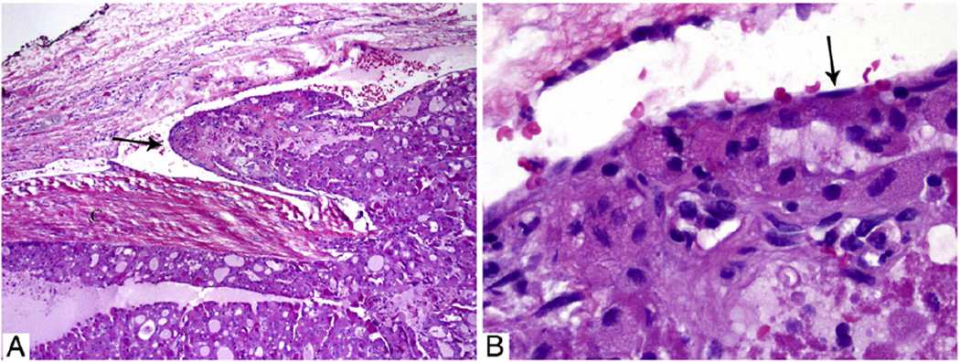
Fig. 2.
Microscopic pictures of a 4.8-cm EHCC with focal VI in a 44-year-old man without distant disease at presentation and not treated by RAI. The patient did not have a recurrence and has no evidence of disease 16 years after diagnosis. A, Medium-power view of the tumor and its capsule with a tumor thrombus hanging (arrow) in the lumen of a vessel located immediately outside the capsule. B, High-power view of the tumor thrombus covered by endothelial cells (arrow).

