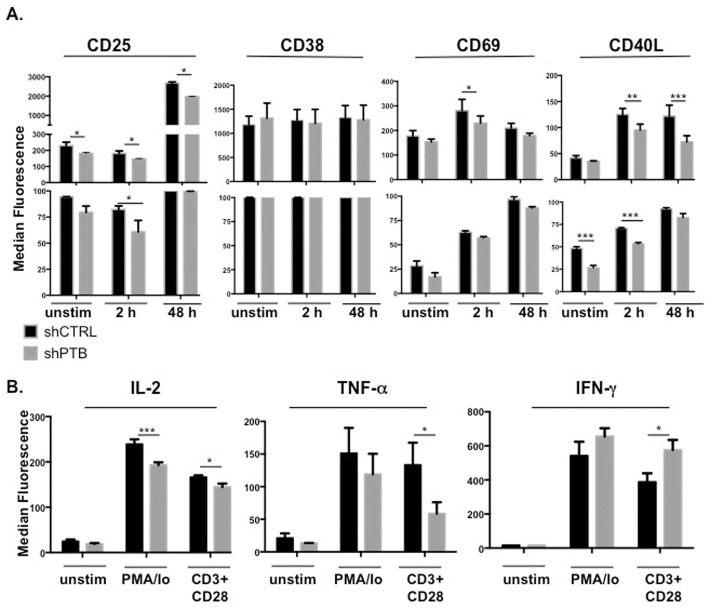
Fig 3. PTB is critical for expression of multiple activation markers.
(A) pLV-shCTRL- or pLV-shPTB-infected CD4 T cells were expanded for 13 days and either left untreated or activated with anti-CD3/anti-CD28 beads for 2 h or 48 h. Following activation, cells were stained with antibodies to selected cell surface markers and analyzed for expression by flow cytometry. Results showing the median fluorescence intensity (top graph) and the percent positive (lower graph) are presented and the boxed bars indicate observed changes in absolute number of positive cells. (B) Expanded CD4 T cells were left untreated or activated with 1 ng/ml PMA and 1 μg/ml Ionomycin for 5 h or anti-CD3/anti-CD28 mAb-bound beads for 48 h and analyzed by intracellular staining with specific antibodies to IL-2, TNFα and IFNγ. Data shown represent the mean and SEM of three independent experiments with *p≤0.05, **p ≤ 0.01, and ***p≤0.005.

