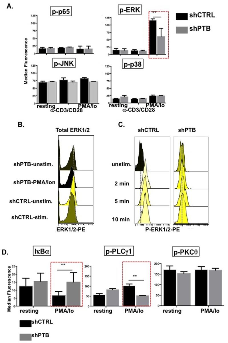
Fig 6. PTBP1 regulates ERK1/2 and NF-κB signaling.
(A) CD4 T cells infected with either pLV-shCTRL or pLV-shPTB lentivirus were activated with either anti-CD3/anti-CD28 beads or 1 ng/ml PMA and 1 μg/ml Io between 2 and 20 min (depending on the optimal response of individual signals). Phosphflow analysis was carried out by fixing cells in paraformaldehyde and permeablizing them for intracellular staining with antibodies to the indicated targets. (B) Histograms showing total (left panel) and phosphorylated ERK1/2 (right panel) in PMA/Io stimulated GFPposCD4pos T cells. (C) Histograms showing ERK1/2 signaling in cells expressing shCTRL and shPTB over 10 min of stimulation with PMA/ionomycin. (D) CD4 T cells infected with either pLV-shCTRL or pLV-shPTB lentivirus were activated with 1 ng/ml PMA and 1 μg/ml Ionomycin for 10 min and analyzed for PLCγ1, PKCθ, and IκBα activity (mean values (+/- SEM) with a *p≤0.05).

