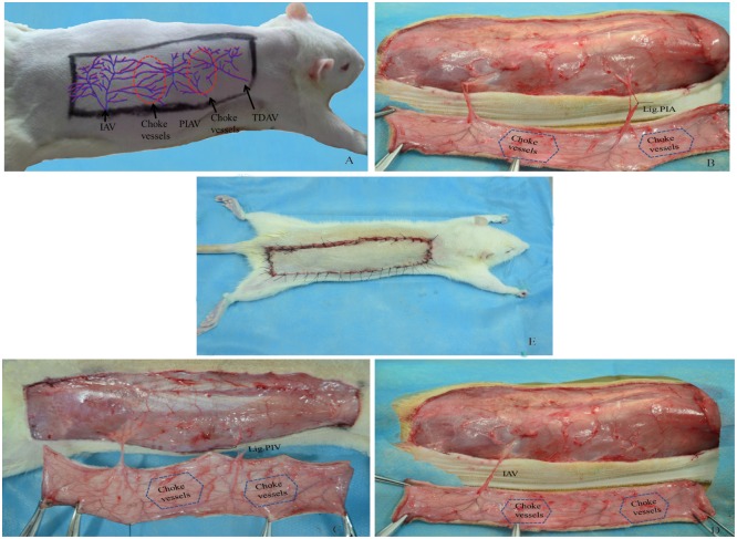
Fig 1. Skin flap design and surgical procedure.
Right dorsal perforator flap model measured 3×12 cm (Fig 1A), Experimental group A, posterior intercostal artery (PIA) was ligated while accompanying vein was preserved (Fig 1B); Experimental group B, posterior intercostal vein (PIV) was ligated while accompanying artery was preserved (Fig 1C); Control group, posterior intercostal artery and vein were ligated, only vascular pedicle (iliolumbar artery and vein, IAV) was preserved (Fig 1D). Finally, the flap was sutured back into its location (Fig 1E). “Choke vessels”: anastomosis area of perforator vessels, IAV: iliolumbar artery and vein, PIAV: posterior intercostal artery and vein, TDAV: thoracodorsal artery and vein.

