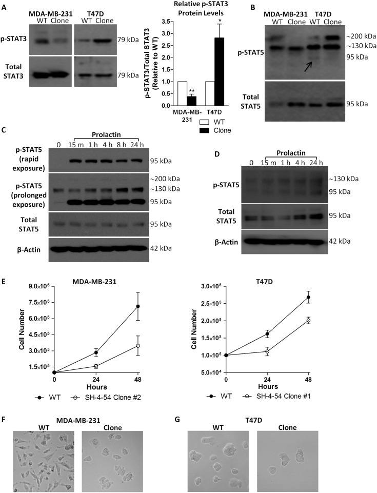
Fig 1.
Chronic treatment of MDA-MB-231 and T47D cells with the STAT3/5 inhibitor SH-4-54, followed by clonal selection and characterization, revealed (A) different levels of phosphorylated STAT3 (p-STAT3) relative to total STAT3 in wild-type (WT) compared to clonally selected MDA-MB-231 and T47D cells, as well as (B) changes in basal levels of phosphorylated STAT5 (p-STAT5) at 95 kDa in T47D cells (indicated by the arrow). The presence of a high molecular weight p-STAT5 band at approximately 200 kDa was also inversely affected in MDA-MB-231 and T47D clonal populations relative to their WT counterparts in a cell-type dependent manner. (C) The ability of T47D cells to undergo rapid and sustained STAT5 activation in response to prolactin treatment was confirmed, with the primary band for p-STAT5 migrating at 95 kDa. (D) Increases in the canonical phosphorylation of STAT5 at 95 kDa could also be detected in MDA-MB-231 cells in response to treatment with prolactin. (E) Less cells were present over 48 hours in SH-4-54-resistant clones compared to MDA-MB-231 and T47D passage-matched WT cells. Representative bright field images (100X magnification, Leica DMIL) shows (F) the characteristic spindle-shaped morphology of WT MDA-MB-231 cells compared to marked morphological changes in SH-4-54 treatment-resistant clones, while (G) no significant changes in morphology were observed between T47D WT and SH-4-54-resistant cells. Data represent the mean of three independent experiments (±SEM) calculated relative to appropriate controls. A star (*) denotes statistically significant differences as determined by a t-test (p<0.05).

