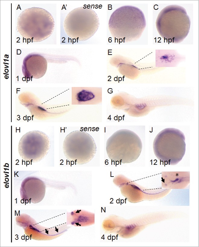
FIGURE 2.

Expression pattern of elovl1a and elovl1b during early development. Expression of elovl1a and elovl1b during embryonic development was analyzed by in situ hybridization. Embryos are presented in animal views (A, A’, H, H’), dorsal views (insets in E, F, L, M) or lateral views (B, C, D, E, F, G, I, J, K, L, M, N). Sense probes for elovl1a (A’) and elovl1b (H’) were used to prove the specificity of antisense probes. Insets in E, F, N, and P are magnified images shown in dorsal views.
