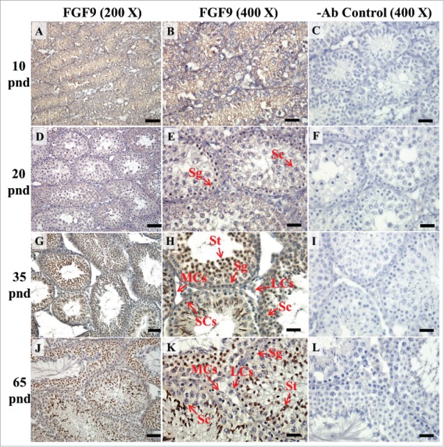
FIGURE 8.

The expression pattern of FGF9 in postnatal mice testis. The expression pattern of FGF9 on postnatal days 10 (A and B), 20 (D and E), 35 (G and H), and 65 (J and K) in mice testis were detected using immunohistochemistry. The cell nucleus was labeled using hematoxylin. Control sections without a primary antibody treatment are arranged on right side of figures 8C, 8F, 8I, and 8L. The long arrow indicates the cell types as illustrated by the abbreviations: LCs, Leydig cells; MCs, myoid cells; SCs, Sertoli cells; Sg, spermatogonia; Sc, spermatocyte; and St, spermatids. Scale bar = 50 µm (200 ×) and 19 µm (400 ×). FGF9 pnd.
