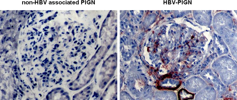
Fig 2. The presence of HBsAg in glomeruli of HBV-PIGN: HbsAg immunostaining was performed with a specific antibody and visualized by the immunoperoxidase method.
Representative pictures show dark red positive stains in glomerulus and in the basal membrane of tubules in HBV-PIGN. The stains are negative in non-HBV associated PIGN.

