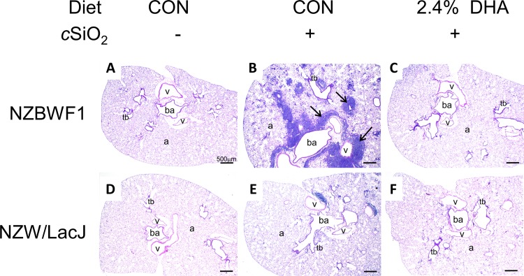
Fig 6. DHA supplementation prevents cSiO2-induced pneumonitis.
Representative photomicrographs of H&E stained lung sections from NZBWF1 (A-C) and NZW/LacJ (D-F) mice exposed to VEH (A, D), cSiO2 fed CON diet (B, E), and cSiO2 fed 2.4% DHA (C, F). Black arrows in light photomicrographs denote marked leukocyte infiltration that circumvented both the vasculature and airways in the lung following cSiO2 exposure (B). Dietary DHA dramatically reduced cSiO2-induced pulmonary inflammation as evident by the absence of cellular accumulation in (C, F). Lymphocytic cell infiltration was semi-quantitatively graded as indicated in Table 3). Abbreviations: ba = bronchiolar airway, v = blood vessel, tb = terminal bronchiole, a = alveolus.

