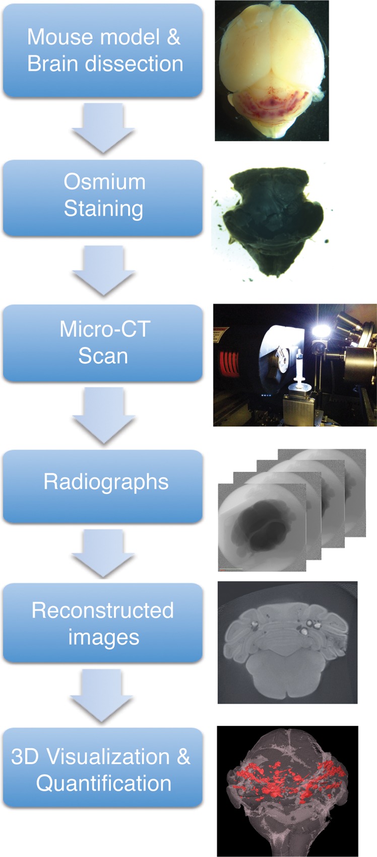
Fig 1. Overview of micro-CT imaging procedure.

Mouse brains were dissected and fixed as described. Following the fixation, only the hindbrain was stained with OsO4 as a contrast staining. Osmium stained hindbrain was then scanned using micro-CT that produces series of radiographs. These radiographs were reconstructed and AVIZO software was used to produce 3D image and quantify lesions in the hindbrain.
