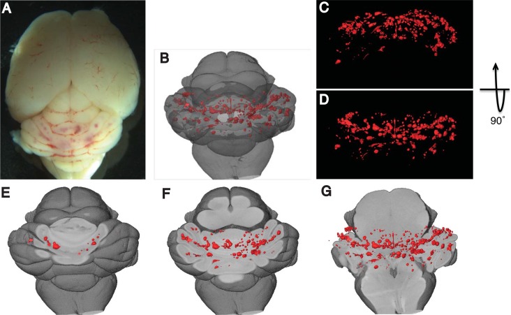
Fig 3. Three-dimensional images of mouse brain with CCM lesions.
A) Macroscopic images of CCM lesions in the hindbrains of Ccm1iECKO mice. B-G) 3-D visualization of CCM lesions at various levels and orientations in the hindbrain. CCM lesions were visualized with the reference to overall hindbrain anatomy (B), from the back (C) and above (D) of hindbrain, and at different depth (E-G) inside hindbrain (red refers to CCM lesions).

