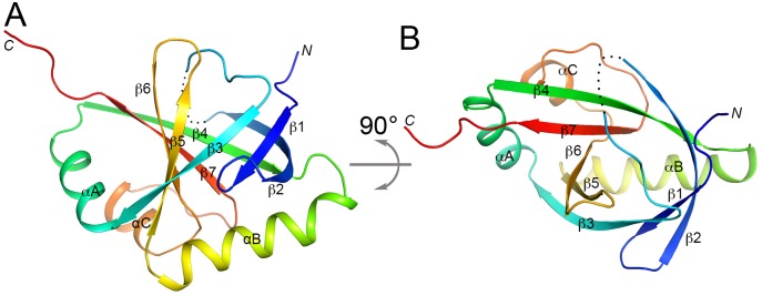
Fig 2. Crystal structure of the SPOC domain of A. thaliana FPA.
(A). Schematic drawing of the structure of FPA SPOC domain, colored from blue at the N terminus to red at the C terminus. The view is from the side of the β-barrel. The disordered segment (residues 460–465) is indicated with the dotted line. (B). Structure of the FPA SPOC domain, viewed from the end of the β-barrel, after 90° rotation around the horizontal axis from panel A. All structure figures were produced with PyMOL (www.pymol.org).

