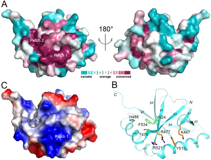
Fig 4. A conserved surface patch of FPA SPOC domain.
(A). Two views of the molecular surface of FPA SPOC domain colored based on sequence conservation among plant FPA homologs. Purple: most conserved; cyan: least conserved. (B). Residues in the conserved surface patch of FPA SPOC domain. The side chains of the residues are shown in stick models, colored orange in the first sub-patch and green in the second. (C). Molecular surface of FPA SPOC domain colored based on electrostatic potential. Blue: positively charged; red: negatively charged.

