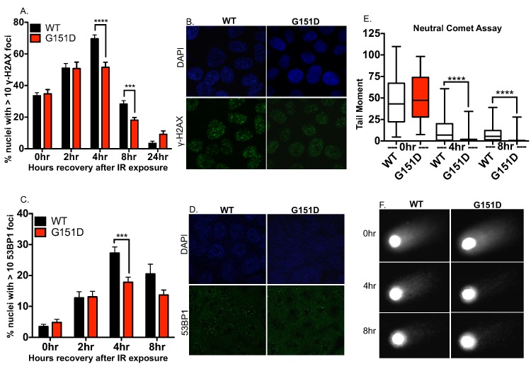
Fig 3. MCF10A cells expressing G151D repair IR-induced double strand breaks more rapidly than WT expressing cells.
A-D. MCF10A pools expressing WT or G151D were exposed to 8GY ionizing radiation, fixed at 0, 2, 4, 8 and 24 (γH2AX only) hours post IR exposure and immunofluorescence was performed. Cells were labeled with a γH2AX antibody (green) (A,B) or 53BP1 antibody (green) (C,D). Labeled cells were visualized using confocal microscopy. A,C. The number of nuclei with >10 foci of γH2AX or 53BP1 was counted. The data are graphed as mean ± SEM (n>500 nuclei) **** p< 0.0001; *** p<0.001. B,D. Representative images of γH2AX foci (B) or 53BP1 (D) at 4 hours post-IR exposure in MCF10A RAD51 WT or RAD51 G151D expressing pools. E, F. MCF10A pools expressing RAD51 WT or G151D were exposed to 8GY ionizing radiation then allowed to recover for 0, 4 and 8 h. Cells were harvested and single cell electrophoresis was performed to quantitate DNA damage using the comet assay. E. Data are graphed as mean ± SEM (number of nuclei counted per group: WT 0hr; 76, G151D 0hr; 80; WT 4hr; 72, G151D 4hr; 97, WT 8hr; 91, G151D 8hr; 100). **** p< 0.0001. F. Representative images from each time point of recovery post IR-exposure.

