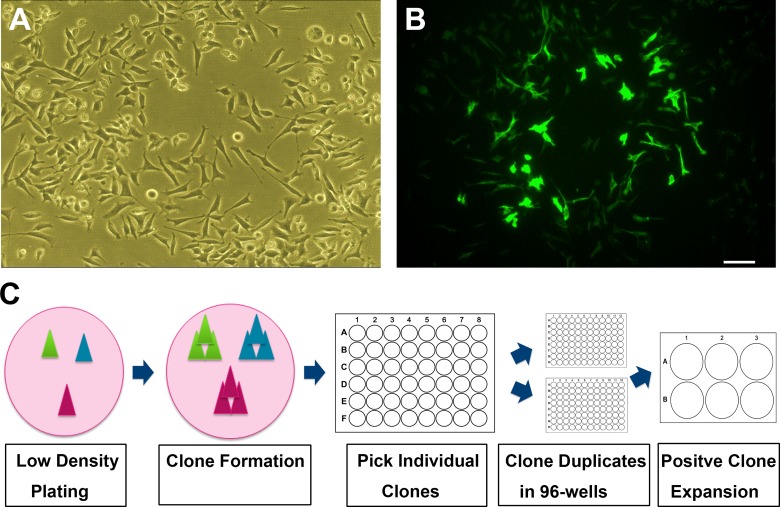
Fig 1. Process of N27 cells clonal purification.
(A): Phase contrast image of unpurified N27 cells grown at low density. (B): Immunostaining for TH with green fluorescence in unpurified N27 cells. Shown is a cluster of cells exhibiting bright TH-positive staining. Most cells shown in phase contrast have no TH-immunoreactivity. (C): Schematic drawing of the clonal culture procedures for purifying N27-A cells. Cells were plated at low density to form individual colonies which were picked up and screened in 48- and 96-well plates. The positive clones were expanded in 6-well plates and 10-cm dishes. Bar, 20 μm for both A and B.

