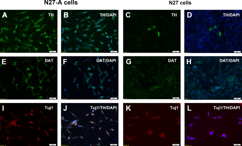
Fig 3. Immunocytochemistry of purified N27-A and unpurified N27 cells for dopamine neuron markers TH and DAT.
The N27 cells were cultured on 8-well chamber slides and immunostained for the dopamine neuron markers TH (A-D) and DAT (E-H). Other wells were double-stained for TH and the neuronal marker Tuj1 (I-L). To image every cell in each well, the nuclear marker DAPI was added to all wells. (A-B): The purified N27-A clone showed strong TH staining in all cells as demonstrated by dual-staining with TH and DAPI. (C-D): The unpurified N27 cell mixture revealed that only a small fraction of the DAPI-labeled cells were TH positive. (E-F): In the purified N27-A clone, all cells had moderate DAT staining as shown with DAT and DAPI double staining. (G-H): In the unpurified N27 cell mixture, very few cells were positive for DAT immunostaining. (I-J): In the purified N27-A clone, all cells were double-positive for Tuj1 and TH. (K-L): While there were few TH-positive cells in the unpurified N27 cell mixture, most cells were Tuj1 positive, demonstrating that the mixed cell population was neuronal. Bar, 50 μm for A-L.

