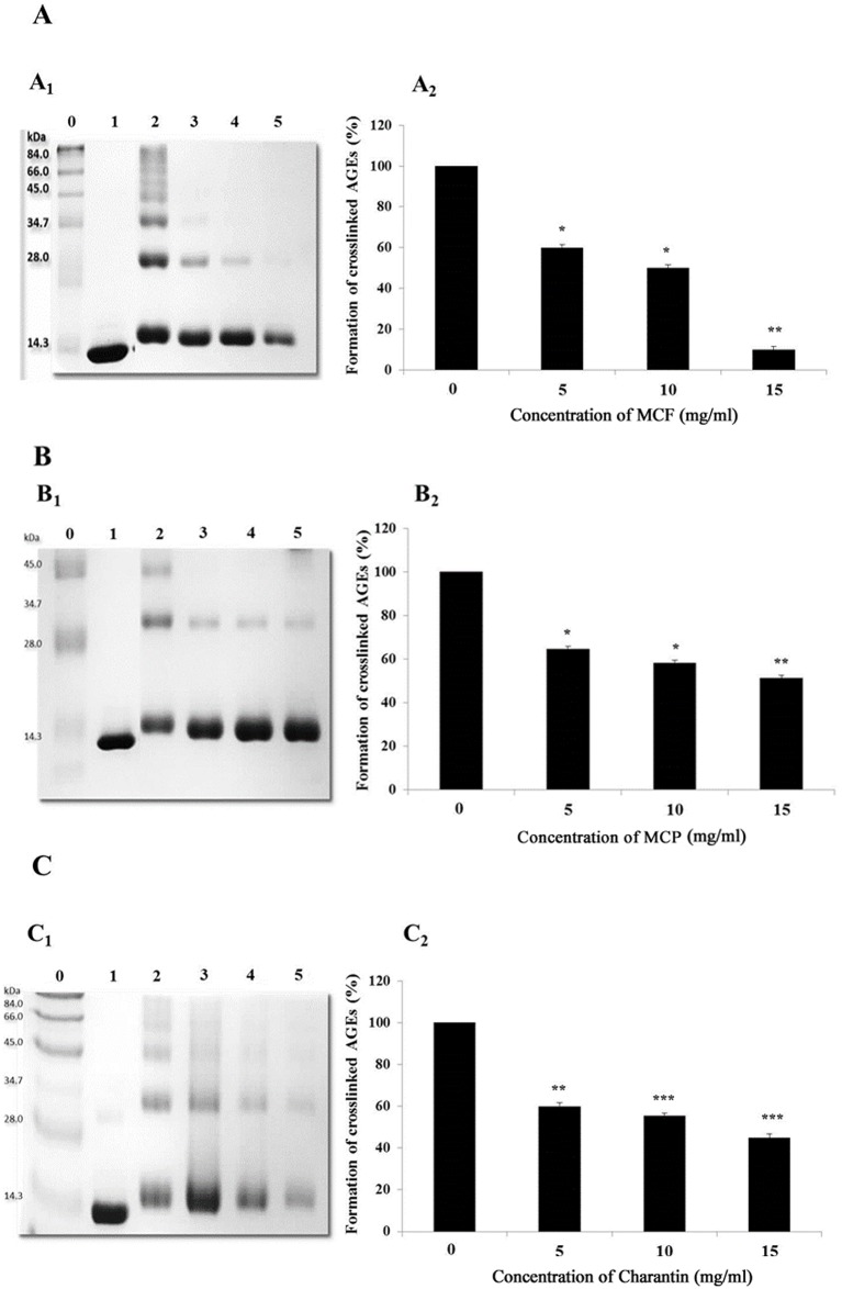
Fig 1. Effects of different concentrations of MCF (A), MCP (B) and charantin (C) on the formation of crosslinked AGEs.
A representative SDS-PAGE gel showing lysozyme (10 mg/ml) incubated alone (lane 1) or in the presence of 0.1 M methylglyoxal (lane 2) in 0.1 M sodium phosphate buffer of pH 7.4 for 3 days at 37°C. The dimer formation resulting from protein crosslinking was determined using different concentrations of the MCF (A1) and MCP (B1) extracts or charantin (C1): 5 mg/ml (lane 3), 10 mg/ml (lane 4) and 15 mg/ml (lane 5). Lane 0 contains the marker proteins. The bar charts show the effects of different concentrations of MCF (A2), MCP (B2) and charantin (C2) on the formation of crosslinked AGEs relative to the control. The results are presented as means ± SDs (n = 3). *: p < 0.05, **: p < 0.01, ***: p < 0.001 vs control.

