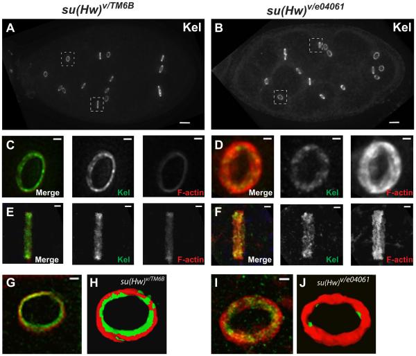
Figure 4.
Rings from su(Hw) mutant egg chambers show significant morphological differences compared to wildtype. Ring canals in wildtype and mutant egg chambers are stained with antibody anti-Kelch in green and Phalloidin in red. Stage 8 egg chambers stained with Kelch are shown in wildtype (A) and su(Hw) mutant (B). Zoom in images of wildtype individual rings from dashed squares in A are shown in C and E. Zoom in images of su(Hw) mutant individual rings from dashed squares in B are shown in D and F. Isosurface images of individual rings in wildtype (G and H) and su(Hw) mutant (I and J) rings were generated using Leica Deblur software, and show the accumulation of actin in rings. The scale bars in egg chamber images represent 10 µm, and in individual ring images represent 1µm.

