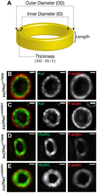
Figure 5.
Ring canals in su(Hw) mutant egg chambers show excess accumulation of structural proteins. A cartoon ring image illustrating the structure and organization of a ring canal (A). Staining of rings at stage 6 using antibodies against ring structural proteins Kelch (B and C), Ovhts-RC (D and E) and F-actin (B-E). Kelch and Ovhts-RC show accumulation at the inner rim in su(Hw) mutants (C and E) but not in wildtype (B and D). Scale bars represent 1µm.

