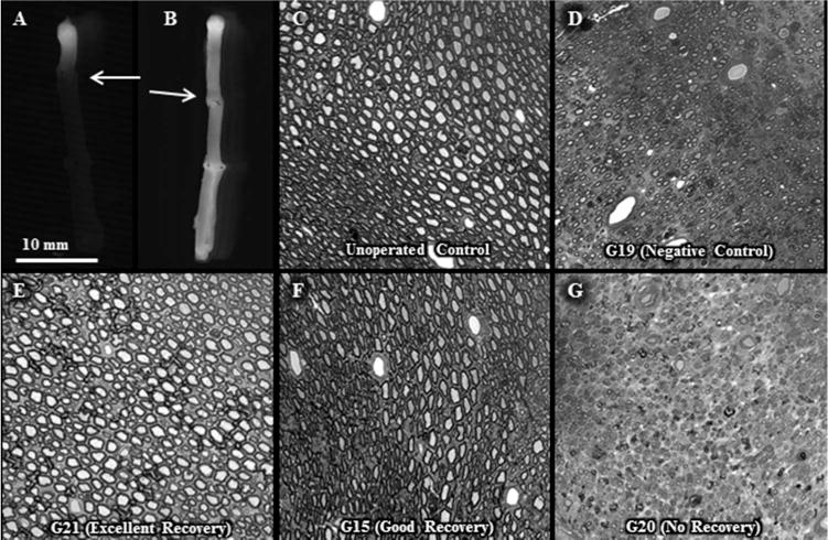
Figure 10. Intra-axonal dye diffusion and axon morphology in allografts.

From Riley et al., 2015) with permission.
A, B. No intra axonal dye diffusion of Texas Red at 1d postoperatively in a negative control allograft (A) but observed for a PEG-fused allograft (B). Arrow: site of proximal cut end of host sciatic nerve micro-sutured to proximal end of donor sciatic allograft.
C–G. Increased myelinated axon viability for PEG-fused allografts compared to negative controls or unsuccessful PEG-fusion at 6w postoperatively. Toluidine-blue, plastic embedded sections of sciatic nerves viewed at 20× for an un-operated control (C) and the mid-allograft region at 6wk postoperatively for a negative control (D) and PEG-fused sciatic nerves showing excellent (E), good (F) or no (G) behavioral recovery as assessed by SFI scores (see Fig. 11 and Riley et al., 2015). Note that successful PEG-fusion is correlated with survival of increased numbers of larger diameter myelinated axons within the allograft segment.
