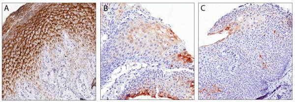
Figure 1. Immunohistochemical staining of SLPI in anal biopsies.
A. Histologically normal squamous epithelium that lines the anus shows strong SLPI staining (brown) in the more differentiated squamous cells in the middle and superficial layers, and less SLPI expression in the basal and stromal layers (40× magnification). B. Epithelium from an AIN1 lesion shows weaker SLPI staining in dysplastic squamous cells (40× magnification). C. Epithelium from an AIN3 lesion shows a further reduction in SLPI staining in dysplastic squamous cells (20× magnification).

