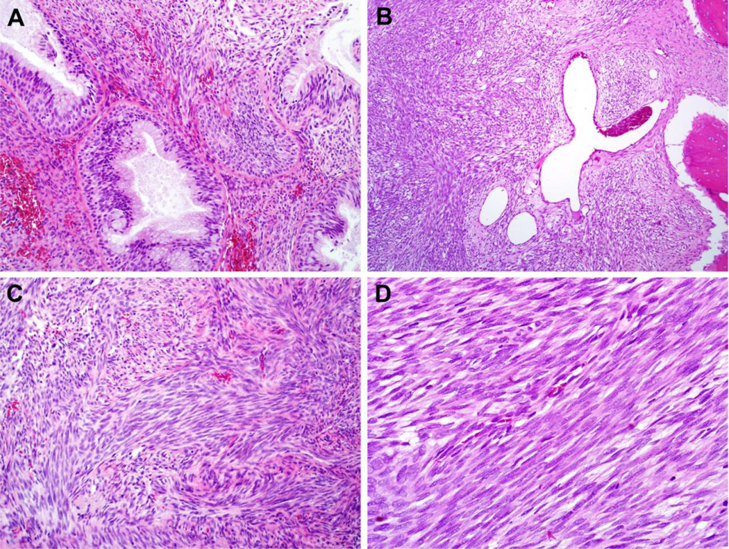
Figure 1.
BSNS frequently demonstrates entrapped benign glands with florid epithelial proliferation (A, X200) as well as prominent staghorn, hemangiopericytoma-like vessels (B, X100). The tumors were composed of dense fascicles of spindle cells with frequent herringbone architecture (C, X200). Tumor cells had a moderate amount of eosinophilic cytoplasm, indistinct cytoplasmic borders, and bland, hypochromatic nuclei with prominent nuclear overlap (D, X400).

