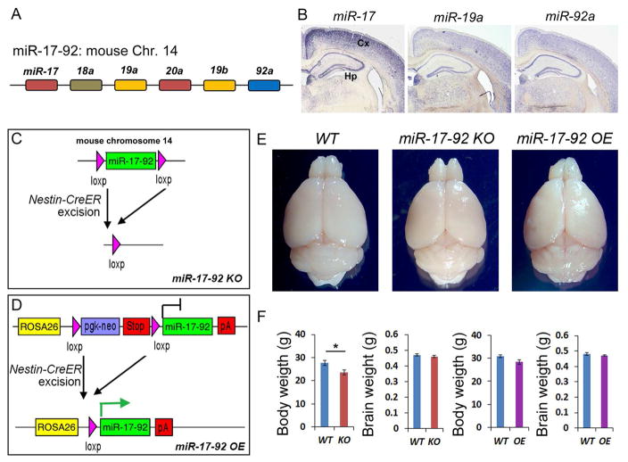
Figure 1. Altering adult hippocampal expression of miR-17-92 in miR-17-92 KO and OE mice.
(A) Schematic genomic organization of miRNAs in the miR-17-92 cluster on mouse chromosome 14. The color code represents miRNAs with the conserved seed sequence.
(B) miR-17 (miR-20a), miR-19a (miR-19b) and miR-92a were expressed in the 12 weeks old adult mouse hippocampus (Hp) and cortex (Cx).
(C and D) Generation of miR-17-92 knockout (KO) and miR-17-92 overexpressing (OE) mice using the Nestin-CreER line.
(E) The whole brain images of wild type (WT), miR-17-92 KO, and miR-17-92 OE mice at the age of 13 weeks old.
(F) The body weight of miR-17-92 KO was slightly reduced, and the body weight of miR-17-92 OE mice was not changed, compared to WT controls. Brain weights of miR-17-92 KO and miR-17-92 OE mice were indistinguishable from WT controls.
Values plotted were means ± s.e.m. n=6 mice per group. *: p < 0.05. A two-tailed, unpaired Student’s t-test was used for comparisons. See also Figures S1 and S2.

