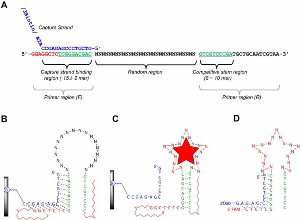
Fig. 1.
An example of SELEX library from Yang et al. [5]. (A) Library structure. This figure shows the relationship between a capture binding region and competitive stem region (green letters in both primer regions) which can induce the structural-switching upon target binding for the partitioning step. (B) An illustration of 2D structure of library binding on the streptavidin coated agarose column. A library element is attached to a column via a capture strand (blue letter in Fig. 1A). The capture strand is attached to an agarose-streptavidin column via a biotinylated oligonucleotide. “B” indicates biotinylation. (C) A library element partitioning mechanism. Upon binding the target, appropriate folding in the random region consequently induces the structural changes, which subsequently leads to dissociation from the capture stand. (D) An example of sensor design. After completion of SELEX, the final sensor is derived from trimming the flanking region of library element.

