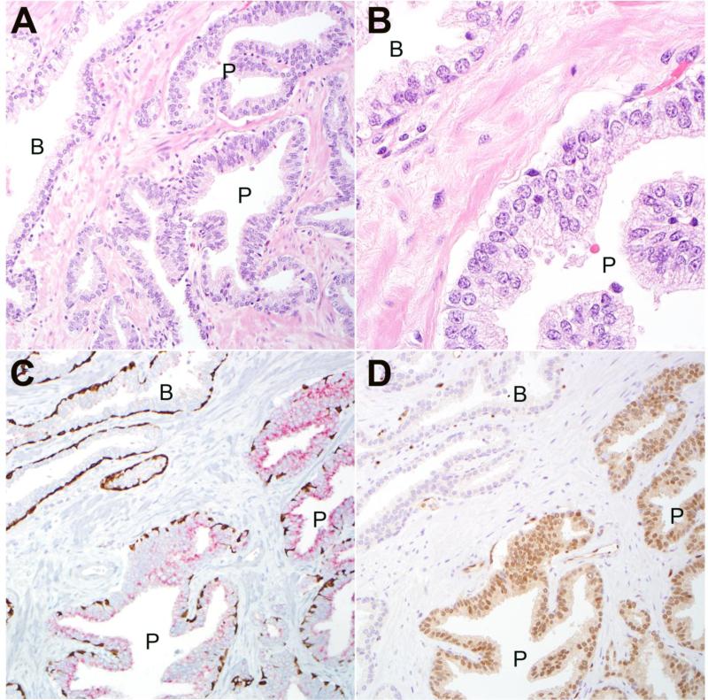
Figure 2.
A: Representative HGPIN lesion (P) from radical cystoprostatectomy with adjacent benign glandular epithelium (B) (200× magnification). B: prominent nucleoli are apparent in HGPIN cells (P) compared to adjacent benign luminal cells (B) (630× magnification). C: Immunostaining with PIN4 cocktail for high molecular weight keratin and p63 demonstrates positively staining basal cells (brown) in both benign (B) and HGPIN (P) glands (200× magnification). Racemase positivity (red) is seen in HGPIN lesion. D: ERG is expressed in nuclei of HGPIN (P) lesion but is negative in adjacent benign (B) glands (200× magnification).

