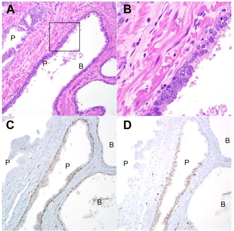
Figure 3.
A: Representative HGPIN lesions (P) from radical cystoprostatectomy with adjacent benign glandular epithelium (B) within the same gland. B: High power image of boxed region from upper left demonstrates prominent nucleoli in HGPIN cells compared to adjacent benign luminal cells (200× magnification) (630× magnification). C: Immunostaining with PIN4 cocktail for high molecular weight keratin and p63 demonstrates positively staining basal cells (brown) in both benign (B) and HGPIN (P) glands. Racemase positivity (red) is absent in this HGPIN lesion (200× magnification). D: Immunostaining for ERG is positive in luminal cells from one of two HGPIN (P) glands and negative and negative in adjacent benign (B) luminal cells within the same gland. An adjacent HGPIN lesion (P) is negative for ERG in the same field (200× magnification).

